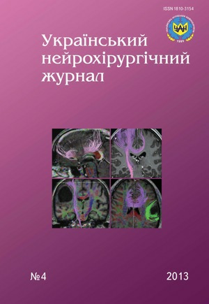
Changes of functionally significant pathways of the brain at low-grade gliomas according to magnetic resonance tractography
DOI:
https://doi.org/10.25305/unj.55411Keywords:
gliomas, MR tractography, pathwaysAbstract
Introduction. The results of magnetic resonance (MR) tractography use in neurosurgical navigation at surgical approach planning and estimation of radicalism of low-grade brain gliomas removing are given.
Materials and methods. Preoperative MR tractography was used in 13 patients, been operated because of low-grade gliomas (with subsequent histological verification).
Results. According to MR tractography low-grade gliomas are characterized by infiltration (in 76.9% cases), destruction of fibers of pathways (in 69.2%), their dislocation (in 23%) due to impact of tumor volume. Surgical tactics at brain gliomas is determined by degree of pathways damage.
Conclusions. MR tractography allows to estimate the degree of pathways damage at brain gliomas, it’s data are used in surgical neuronavigation.References
1. Kulikov KA. Dostovernyye kriterii prognoza vyzhivaniya bol'nykh tserebral'noy gliomoy posle kombinirovannoy terapii [Significant criteria for survival prognosis of patients with cerebral glioma after combination therapy]. Perm Med Zhurn. 2012;29(2):31-7. Russian.
2. Zozulia IuA, Patsko IaV, Nikiforova AN. [Epidemiological studies in neuro-oncology: the state of the art in Ukraine and abroad]. Zh Vopr Neirokhir Im N N Burdenko. 1998 Jul-Sep;(3):50-4. Russian. [PubMed]
3. Rozumenko VD. [Epidemiology of brain tumors: factors of statistics]. Ukr Neurosurg J. 2002;3:47-8. Russian.
4. Christoforidis GA, Yang M, Abduljalil A, Chaudhury AR, Newton HB, McGregor JM, Epstein CR, Yuh WT, Watson S, Robitaille PM. "Tumoral pseudoblush" identified within gliomas at high-spatial-resolution ultrahigh-field-strength gradient-echo MR imaging corresponds to microvascularity at stereotactic biopsy. Radiology.2012 Jul;264(1):210-7. [CrossRef] [PubMed]
5. Sanai N, Berger MS. Extent of resection influences outcomes for patients with gliomas. Rev Neurol (Paris). 2011 Oct;167(10):648-54. [CrossRef] [PubMed]
6. Rozumenko VD., Chuvashova OYu., Ruditsa VI, Rozumenko AV. [MR-tractography in image-guide surgery of brain tumors]. Ukrainian Neurosurgical Journal. 2011;2:65-8. Russian.
7. Duffau H. New concepts in surgery of WHO grade II gliomas: functional brain mapping, connectionism and plasticity - a review. J Neurooncol. 2006 Aug;79(1):77-115. [CrossRef] [PubMed]
8. Hendler T, Pianka P, Sigal M, Kafri M, Ben-Bashat D, Constantini S, Graif M, Fried I, Assaf Y. Delineating gray and white matter involvement in brain lesions: three-dimensional alignment of functional magnetic resonance and diffusion-tensor imaging. J Neurosurg. 2003 Dec;99(6):1018-27.[CrossRef] [PubMed]
9. Lee SK, Kim DI, Kim J, Kim DJ, Kim HD, Kim DS, Mori S. Diffusion-tensor MR imaging and fiber tractography: a new method of describing aberrant fiber connections in developmental CNS anomalies. Radiographics. 2005 Jan-Feb;25(1):53-68. [CrossRef]
10. Mori S, Frederiksen K, van Zijl PC, Stieltjes B, Kraut MA, Solaiyappan M, Pomper MG. Brain white matter anatomy of tumor patients evaluated with diffusion tensor imaging. Ann Neurol. 2002 Mar;51(3):377-80. [CrossRef] [PubMed]
11. Holodny AI, Schwartz TH, Ollenschleger M, Liu WC, Schulder M. Tumor involvement of the corticospinal tract: diffusion magnetic resonance tractography with intraoperative correlation. J Neurosurg. 2001 Dec;95(6):1082. [CrossRef] [PubMed]
12. Bozzali M, MacPherson SE, Cercignani M, Crum WR, Shallice T, Rees JH. White matter integrity assessed by diffusion tensor tractography in a patient with a large tumor mass but minimal clinical and neuropsychological deficits. Funct Neurol. 2012 Oct-Dec;27(4):239-46. [PubMed]
Downloads
Published
How to Cite
Issue
Section
License
Copyright (c) 2013 Olga Chuvashova, Kristiana Robаk

This work is licensed under a Creative Commons Attribution 4.0 International License.
Ukrainian Neurosurgical Journal abides by the CREATIVE COMMONS copyright rights and permissions for open access journals.
Authors, who are published in this Journal, agree to the following conditions:
1. The authors reserve the right to authorship of the work and pass the first publication right of this work to the Journal under the terms of Creative Commons Attribution License, which allows others to freely distribute the published research with the obligatory reference to the authors of the original work and the first publication of the work in this Journal.
2. The authors have the right to conclude separate supplement agreements that relate to non-exclusive work distribution in the form of which it has been published by the Journal (for example, to upload the work to the online storage of the Journal or publish it as part of a monograph), provided that the reference to the first publication of the work in this Journal is included.


