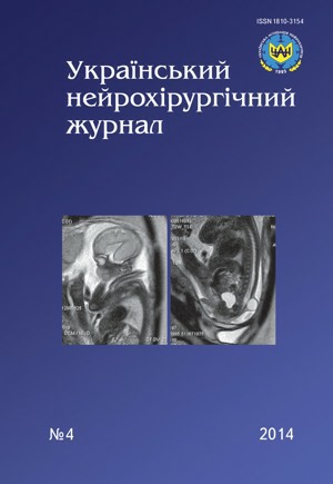
Role of magnetic resonance imaging in prenatal diagnostics of congenital malformations of central nervous system
DOI:
https://doi.org/10.25305/unj.46600Keywords:
pregnancy, fetus, CNS pathology, MRIAbstract
Introduction. The opportunities, safety and role of magnetic resonance imaging (MRI) in diagnostics of congenital malformations of central nervous system (CNS) are given on the base of the literature analysis and results of own observations, the need of MRI inclusion into protocol/algorithm of inspection of pregnant women in Ukraine is shown at detection of any ultrasound changes for post-natal treatment planning, including neurosurgical interventions.
Materials and method of the study. Features of MRI program, used for fetus examing, time of the procedure are described on the base of literature analysis and own observations.
Results. Methods of MRI study of fetal CNS are described: brain biometrics, condition of brain ventricles, analysis of gyrus and sulcus development, migration of neurons and white and gray substance of a brain forming, study of a spine.
According to results of own observations, the possibility of ultrasound and MRI diagnoses divergence was shown, most accurate other data at MRI.
Conclusion. More than 25 years of international experience of MRI in prenatal diagnostics at congenital malformations assumption according to ultrasound data, especially of CNS, and results of own study confirm high informative value and safety of the method, high specificity of characteristics of pathological changes, indicating the need to introduce MRI in the algorithm of examination of pregnant women in Ukraine.
References
Barashnev YU, Bakharev YU, Novikov P. Diagnostika i lecheniye vrozhdennykh i nasledstvennykh zabolevaniy u detey. Moscow: Triada-KH; 2004. Russian.
Polunin V, Nesterenko Ye, Popov V., Solomatin D. Mediko-sotsial'nyye faktory riska vozniknoveniya porokov razvitiya spinnogo mozga. Ros Med Zhurn. 2006;1:3–6. Russian.
Bokeriya L, Stupakov I, Zaychenko N, Gudukova R. Vrozhdennyye anomalii (poroki razvitiya) v Rossiyskoy Federatsii. Det Bolnitsa. 2003;1:7–14. Russian.
Banovic J, Banovic V, Roje D. The influence of drug abuse on perinatal outcome. Proceedings of the 5th World Congress of Perinatal Medicine. Barcelona, 2001. p.504–507.
Kashina Ye.V. Kliniko-morfologicheskiye osobennosti vrozhdennykh porokov razvitiya tsentral'noy nervnoy sistemy v ontogeneze u detey. [Clinico-morphological features of congenital malformations of the central nervous system in the ontogenesis of children]. [dissertation]. Khabarovsk. (Russia) State. institution of higher prof. Education "Vladivostok State Medical University, Federal Agency for Health and Social Development." 2008. Russian.
Makogon A, Makhotin A, Korostyshevskaya A, Kalenitskaya L. Patologiya mozolistogo tela. Trekhmernaya ul'trazvukovaya diagnostika i MR-tomografiya. Ul'trazvuk i Funkts. Diagnostika. 2008;2:79. Russian.
Wilhelm C, Keck C, Hess S, Korinthenberg R, Breckwoldt M. Ventriculomegaly Diagnosed by Prenatal Ultrasound and Mental Development of the Children. Fetal Diagnosis and Therapy. 1998;13(3):162-166. CrossRef
Korniyenko V, Pronin I. Diagnosticheskaya neyroradiologiya. Moscow:IP «Andreyeva T.M.»; 2006. Russian.
Yudina Ye, Medvedev M. Osnovy prenatal'noy diagnostiki. Moscow:RAVUZDPAG, Real'noye vremya; 2002. Russian.
Yusupov K, Ibatulin M, Mikhaylov I, Panov V. MRT v antenatal'noy diagnostike anomaliy vnutriutrobnogo ploda. Radiologiya — Praktika. 2006;2:24–42. Russian.
Smith F.W. NMR imaging in pregnancy. The Lancet. 1983;321(8314-8315):61-62. CrossRef
Thickman D, Mintz M, Mennuti M, Kressel H. MR Imaging of Cerebral Abnormalities In Utero. Journal of Computer Assisted Tomography. 1984;8(6):1058-1061. CrossRef
Glenn OA, Barkovich AJ. Magnetic resonance imaging of fetal brain and spine: an increasingly important tool in prenatal diagnosis, part 1. AJNR Am J Neuroradiol. 2006;27(8):1604-1611. PubMed
Glenn OA, Barkovich J. Magnetic resonance imaging of fetal brain and spine: an increasingly important tool in prenatal diagnosis, part 2. AJNR Am J Neuroradiol. 2006 Oct;27(9):1807-1814. Review. PubMed
Coakley F, Glenn O, Qayyum A, Barkovich A, Goldstein R, Filly R. Fetal MRI: A Developing Technique for the Developing Patient. American Journal of Roentgenology. 2004;182(1):243-252. CrossRef
Robinson I. Fetal magnetic resonance imaging: a valuable diagnostic tool. Infant. 2009;5(4):124–126.
Korostyshevskaya AM. Magnitno-rezonansnaya tomografiya v diagnostike anomaliy sredinnykh struktur golovnogo mozga u ploda. Med Visualizatsiya. 2010;4:112–118.Russian.
Trofimova T, Khalikov A, Voronin D, Pavlova N. Nuzhna li prenatal'naya magnitno-rezonansnaya tomografiya? Luchevaya diagnostika i terapiya. 2011;2:13–21.Russian.
Rogozhyn V, Rozhkova Z, Kirillova L. MRI and 1H MRS for early detection of hypoxic injury of the fetal brain. Eur. J. Neuroradiol. 2002;11(3):352.
Rogozhyn V, Rozhkova Z, Kirillova L, Lukjanova E, Perfilov A. Early detection of the hypoxic injury of the fetal human brain by using MRI and 1H MRS. Brain and Development. 2002;24(6):589–590.
Kyrylova L, Rohozhyn V, Perfilov O, Rozhkova Z, Myronyak L. Suchasni diahnostychni mozhlyvosti neyrovizualizatsiyi u perynatolohiyi. Pediatriya, akusherstvo ta hinekolohiya. 2003;2 addition:52–53. Ukrainian.
Kyrylova L, Rohozhyn V, Pysareva S, Rozhkova Z, Myronyak L, Ryabikin O. Diahnostychna otsinka stanu TSNS ploda, novonarodzhenykh i ditey rannoho viku metodamy suchasnoyi neyrovizualizatsiyi (MRT i MRS). Pediatriya, akusherstvo ta hinekolohiya. 2003;(3 Suppl):3–6. Ukrainian.
Rogozhyn V, Rozhkova Z, Kirillova L. MRI and 1H MRS for monitoring of the fetal brain development. Eur Radiol. 2003;13(1):285.
Rogozhyn V, Rozhkova Z. MRI and MRS of the human fetal brain. Radiology. 2003;223(2):396.
Myronyak L, Rogozhyn V, Ryabikin O, Kirillova L. MRI in detection of fetal brain anomalies. Montreal:Book of abstracts, 23-rd ICR; 2004.
Rogozhin V. MRT v ginekologicheskoy praktike. R.E.J.R. 2012;2(3):27–40. Russian.
Rodrigues M, Vega Fernandez V, Ten P, Pedregosa J, Fernandez – Mayoralas D, de la Pena M. Fetal MRI in CNC abnormalities. Relevant issues for obstetriciens. R.A.R. 2010;74(4):1–14.
Girard N, Raybaud C, Gambarelli D, Figarella-Branger D. Fetal brain MR imaging. Magn Reson Imaging Clin N Am. 2001 Feb;9(1):19-56, vii. PubMed
Weinreb J, Lowe T, Cohen J, Kutler M. Human fetal anatomy: MR imaging. Radiology. 1985;157(3):715-720. CrossRef
Powell MC, Worthington BS, Buckley JM, Symonds EM. Magnetic resonance imaging (MRI) in obstetrics. II. Fetal anatomy. BJOG: An International Journal of Obstetrics and Gynaecology. 1988;95(1):38-46. CrossRef
Salomon L, Siauve N, Balvay D et al. Placental Perfusion MR Imaging with Contrast Agents in a Mouse Model1. Radiology. 2005;235(1):73-80. CrossRef
Grobner T, Prischl F. Gadolinium and nephrogenic systemic fibrosis. Kidney International. 2007;72(3):260-264. CrossRef
Saleem S. Fetal MRI: An approach to practice: A review. Journal of Advanced Research. 2014;5(5):507-523. CrossRef
Agid R, Lieberman S, Nadjari M, Gomori J. Prenatal MR diffusion-weighted imaging in a fetus with hemimegalencephaly. Pediatr Radiol. 2005;36(2):138-140. CrossRef
Baldoli C, Righini A, Parazzini C, Scotti G, Triulzi F. Demonstration of acute ischemic lesions in the fetal brain by diffusion magnetic resonance imaging. Annals of Neurology. 2002;52(2):243-246. CrossRef
Brugger P, Stuhr F, Lindner C, Prayer D. Methods of fetal MR: beyond T2-weighted imaging. European Journal of Radiology. 2006;57(2):172-181. CrossRef
Baker P, Johnson I, Harvey P, Gowland P, Mansfield P. A three-year follow-up of children imaged in utero with echo-planar magnetic resonance. American Journal of Obstetrics and Gynecology. 1994;170(1):32-33. CrossRef
Glenn O, Coakley F. MRI of the Fetal Central Nervous System and Body. Clinics in Perinatology. 2009;36(2):273-300. CrossRef
Korostyshevskaya A, Savelov A. Rol' magnitno-rezonansnoy tomografii ploda v diagnostike vrozhdennykh porokov razvitiya. Byul Cib Med. 2012;5:128–132. Russian.
Garel C. The role of MRI in the evaluation of the fetal brain with an emphasis on biometry, gyration and parenchyma. Pediatr Radiol. 2004;34(9). CrossRef
Viñals F, Muñoz M, Naveas R, Shalper J, Giuliano A. The fetal cerebellar vermis: anatomy and biometric assessment using volume contrast imaging in the C-plane (VCI-C). Ultrasound in Obstetrics and Gynecology. 2005;26(6):622-627. CrossRef
Garel C, Alberti C. Coronal measurement of the fetal lateral ventricles: comparison between ultrasonography and magnetic resonance imaging. Ultrasound in Obstetrics and Gynecology. 2005;27(1):23-27. CrossRef
Bowerman RA. Normal fetal anatomic survey. In: Nyberg D, Gahan J, Pretorius D, Pilu G. eds. Diagnostic imaging of fetal anomalies. Philadelphia:Lippincott Williams & Wilkins; 2003.
Brisse H, Fallet C, Sebag G, Nessmann C, Blot P, Hassan M. Supratentorial parenchyma in the developing fetal brain: in vitro MR study with histologic comparison. AJNR Am J Neuroradiol. 1997 Sep;18(8):1491-7. PubMed
2. Chung H, Chen C, Zimmerman R, Lee K, Lee C, Chin S. T2-Weighted Fast MR Imaging with True FISP Versus HASTE. American Journal of Roentgenology. 2000;175(5):1375-1380. CrossRef
Brace V, Grant S, Brackley K, Kilby M, Whittle M. Prenatal diagnosis and outcome in sacrococcygeal teratomas: a review of cases between 1992 and 1998. Prenatal Diagnosis. 2000;20(1):51-55. CrossRef
Downloads
Published
How to Cite
Issue
Section
License
Copyright (c) 2014 Olga Chuvashova

This work is licensed under a Creative Commons Attribution 4.0 International License.
Ukrainian Neurosurgical Journal abides by the CREATIVE COMMONS copyright rights and permissions for open access journals.
Authors, who are published in this Journal, agree to the following conditions:
1. The authors reserve the right to authorship of the work and pass the first publication right of this work to the Journal under the terms of Creative Commons Attribution License, which allows others to freely distribute the published research with the obligatory reference to the authors of the original work and the first publication of the work in this Journal.
2. The authors have the right to conclude separate supplement agreements that relate to non-exclusive work distribution in the form of which it has been published by the Journal (for example, to upload the work to the online storage of the Journal or publish it as part of a monograph), provided that the reference to the first publication of the work in this Journal is included.


