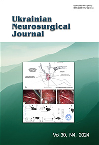
Minimizing skull defects in retrosigmoid approach: precision mapping of the sigmoid sinus with mastoid emissary vein canal
DOI:
https://doi.org/10.25305/unj.313077Keywords:
Mastoid emissary vein canal, retrosigmoid craniotomy, mastoid foramen, cranial topographyAbstract
Objective. The retrosigmoid approach is a commonly used cranial approach to the cerebellopontine angle lesions, vascular and nerve pathologies. This study aims to develop a practical technique for intraoperative mapping of the sigmoid sinus using the topography of the mastoid emissary vein (MEV) canal to improve the accuracy of retrosigmoid craniotomy, and minimize postoperative adverse outcomes.
Materials and methods. Consecutive patients who underwent retrosigmoid approaches for cerebellopontine angle occupying lesions from October 2023 through August 2024 were included in the study. Perioperative computed tomography (CT) was performed with a slice thickness 0.5 mm in the axial plane. The projection of the internal opening of the MEV canal onto the external surface of the mastoid process was determined as the posterior border sigmoid sinus and anterior border for craniotomy. Comparative analyses were performed using t-test and Chi-square test.
Results. A total of 20 patients were operated for neoplasms occupying the cerebellopontine angle using retrosigmoid approach. The average measured distance from the external opening of the MEV canal to the projection of sigmoid sinus posterior border was 9.36 ± 2.17 mm (range 6.3–13.20 mm). The postoperative CT data showed statistically significant differences between the study and control groups in measures of bone window (p = 0.057) and surrounding cranial defect (p < 0.001). The size of bone flaps was slightly similar in all groups (p = 0.114). The mean cranial defect in the study group was almost twice smaller than in the control group 22.4% vs. 44.5% respectively.
Conclusions. This study confirms the utility of mastoid emissary vein canal topography in improving the accuracy of retrosigmoid craniotomy. By facilitating precise sigmoid sinus mapping, the technique reduces the extent of bone removal and minimizes postoperative cranial defect.
References
1. Sepehrnia A, Knopp U. Osteoplastic lateral suboccipital approach for acoustic neuroma surgery. Neurosurgery. 2001;48(1):229-30; discussion 230-1. [CrossRef] [PubMed]
2. Zhou W, Di G, Rong J, Hu Z, Tan M, Duan K, Jiang X. Clinical applications of the mastoid emissary vein. Surg Radiol Anat. 2023 Jan;45(1):55-63. [CrossRef] [PubMed] [PubMed Central]
3. Chaiyamoon A, Schneider K, Iwanaga J, Donofrio CA, Badaloni F, Fioravanti A, Tubbs RS. Anatomical study of the mastoid foramina and mastoid emissary veins: classification and application to localizing the sigmoid sinus. Neurosurg Rev. 2023 Dec 19;47(1):16. [CrossRef] [PubMed]
4. Hampl M, Kachlik D, Kikalova K, Riemer R, Halaj M, Novak V, Stejskal P, Vaverka M, Hrabalek L, Krahulik D, Nanka O. Mastoid foramen, mastoid emissary vein and clinical implications in neurosurgery. Acta Neurochir (Wien). 2018 Jul;160(7):1473-1482. [CrossRef] [PubMed]
5. Raso JL, Gusmão SN. A new landmark for finding the sigmoid sinus in suboccipital craniotomies. Neurosurgery. 2011 Mar;68(1 Suppl Operative):1-6; discussion 6. [CrossRef] [PubMed]
6. Kubo M, Mizutani T, Shimizu K, Matsumoto M, Iizuka K. New methods for determination of the keyhole position in the lateral suboccipital approach to avoid transverse-sigmoid sinus injury: Proposition of the groove line as a new surgical landmark. Neurochirurgie. 2021 Jul;67(4):325-329. [CrossRef] [PubMed]
7. Hamasaki T, Morioka M, Nakamura H, Yano S, Hirai T, Kuratsu J. A 3-dimensional computed tomographic procedure for planning retrosigmoid craniotomy. Neurosurgery. 2009 May;64(5 Suppl 2):241-5; discussion 245-6. [CrossRef] [PubMed]
8. Tubbs RS, Loukas M, Shoja MM, Bellew MP, Cohen-Gadol AA. Surface landmarks for the junction between the transverse and sigmoid sinuses: application of the "strategic" burr hole for suboccipital craniotomy. Neurosurgery. 2009 Dec;65(6 Suppl):37-41; discussion 41. [CrossRef] [PubMed]
9. da Silva EB Jr, Leal AG, Milano JB, da Silva LF Jr, Clemente RS, Ramina R. Image-guided surgical planning using anatomical landmarks in the retrosigmoid approach. Acta Neurochir (Wien). 2010 May;152(5):905-10. [CrossRef] [PubMed]
10. Hu W, Zhou J, Liu ZM, Li Y, Cai QF, Hu XM, Yu YB. Computed Tomography Study of the Retrosigmoid Craniotomy Keyhole Approach Using Surface Landmarks. Int J Clin Pract. 2023 Mar 1;2023:5407912. [CrossRef] [PubMed] [PubMed Central]
11. Reis CV, Deshmukh V, Zabramski JM, Crusius M, Desmukh P, Spetzler RF, Preul MC. Anatomy of the mastoid emissary vein and venous system of the posterior neck region: neurosurgical implications. Neurosurgery. 2007 Nov;61(5 Suppl 2):193-200; discussion 200-1. [CrossRef] [PubMed]
12. Roser F, Ebner FH, Ernemann U, Tatagiba M, Ramina K. Improved CT Imaging for Mastoid Emissary Vein Visualization Prior to Posterior Fossa Approaches. J Neurol Surg A Cent Eur Neurosurg. 2016 Nov;77(6):511-514. [CrossRef] [PubMed]
13. Hall S, Peter Gan YC. Anatomical localization of the transverse-sigmoid sinus junction: Comparison of existing techniques. Surg Neurol Int. 2019 Sep 27;10:186. [CrossRef] [PubMed] [PubMed Central]
14. Choque-Velasquez J, Hernesniemi J. One burr-hole craniotomy: Upper retrosigmoid approach in helsinki neurosurgery. Surg Neurol Int. 2018 Aug 14;9:163. [CrossRef] [PubMed] [PubMed Central]
Published
How to Cite
Issue
Section
License
Copyright (c) 2024 Artem V. Rozumenko, Mykola V. Yehorov, Vasyl V. Shust, Dmytro M. Tsiurupa, Anton M. Dubrovka, Petro M. Onishchenko, Volodymyr O. Fedirko

This work is licensed under a Creative Commons Attribution 4.0 International License.
Ukrainian Neurosurgical Journal abides by the CREATIVE COMMONS copyright rights and permissions for open access journals.
Authors, who are published in this Journal, agree to the following conditions:
1. The authors reserve the right to authorship of the work and pass the first publication right of this work to the Journal under the terms of Creative Commons Attribution License, which allows others to freely distribute the published research with the obligatory reference to the authors of the original work and the first publication of the work in this Journal.
2. The authors have the right to conclude separate supplement agreements that relate to non-exclusive work distribution in the form of which it has been published by the Journal (for example, to upload the work to the online storage of the Journal or publish it as part of a monograph), provided that the reference to the first publication of the work in this Journal is included.


