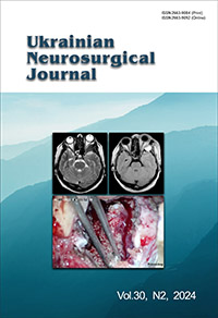
Minimally invasive orbito-zygomatic access for cranio-orbital hyperostotic meningiomas. Case report
DOI:
https://doi.org/10.25305/unj.298906Keywords:
meningioma, hyperostotic lesion, orbit, minimally invasive surgeryAbstract
Application into clinical practice of a minimally invasive surgical approach to the removal of hyperostotic cranioorbital meningiomas.
This publication is based on the analysis of a clinical case of 49-year-old woman with exophthalmos, and the absence of neurological deficits. A non-standard approach to remove a cranio-orbital hyperostotic meningioma through a minimally invasive orbito-zygomatic approach was used.
The main principle of proposed surgical approach was to remove first the hyperostosis, followed by the areas of dura mater involved by the tumor, according to the "outside-in" principle. According to the intraoperative process and the results of MRI control, it was possible to achieve total removal of both the affected dura mater and the hyperostotic lesion.
The minimally invasive transorbital approach opens a wide corridor for surgery of the para and retroorbital space and allows using the "outside-in" method, to remove not only hyperostosis but also the area of damage to the dura mater.
References
1. Wiemels J, Wrensch M, Claus EB. Epidemiology and etiology of meningioma. J Neurooncol. 2010 Sep;99(3):307-14. [CrossRef] [PubMed] [PubMed Central]
2. Cao J, Yan W, Li G, Zhan Z, Hong X, Yan H. Incidence and survival of benign, borderline, and malignant meningioma patients in the United States from 2004 to 2018. Int J Cancer. 2022 Dec 1;151(11):1874-1888. [CrossRef] [PubMed]
3. Simas NM, Farias JP. Sphenoid Wing en plaque meningiomas: Surgical results and recurrence rates. Surg Neurol Int. 2013 Jul 9;4:86. [CrossRef] [PubMed] [PubMed Central]
4. Meningiomas. Their Classification, Regional Behaviour, Life History, and Surgical End Results. Bull Med Libr Assoc. 1938 Dec;27(2):185. [PubMed Central]
5. Cushing H. The cranial hyperostoses produced by meningeal endotheliomas. Archives of Neurology And Psychiatry. 1922 Aug 1;8(2):139. [CrossRef]
6. Bikmaz K, Mrak R, Al-Mefty O. Management of bone-invasive, hyperostotic sphenoid wing meningiomas. J Neurosurg. 2007 Nov;107(5):905-12. [CrossRef] [PubMed]
7. Honeybul S, Neil-Dwyer G, Lang DA, Evans BT, Ellison DW. Sphenoid wing meningioma en plaque: a clinical review. Acta Neurochir (Wien). 2001 Aug;143(8):749-57; discussion 758. [CrossRef] [PubMed]
8. Pieper DR, Al-Mefty O, Hanada Y, Buechner D. Hyperostosis associated with meningioma of the cranial base: secondary changes or tumor invasion. Neurosurgery. 1999 Apr;44(4):742-6; discussion 746-7. [CrossRef] [PubMed]
9. Carrizo A, Basso A. Current surgical treatment for sphenoorbital meningiomas. Surg Neurol. 1998 Dec;50(6):574-8. [CrossRef] [PubMed]
10. Schepers S, Ioannides C, Fossion E. Surgical treatment of exophthalmos and exorbitism: a modified technique. J Craniomaxillofac Surg. 1992 Oct;20(7):313-6. [CrossRef] [PubMed]
11. Talacchi A, De Carlo A, D'Agostino A, Nocini P. Surgical management of ocular symptoms in spheno-orbital meningiomas. Is orbital reconstruction really necessary? Neurosurg Rev. 2014 Apr;37(2):301-9; discussion 309-10. [CrossRef] [PubMed]
12. Gorman L, Ruben J, Myers R, Dally M. Role of hypofractionated stereotactic radiotherapy in treatment of skull base meningiomas. J Clin Neurosci. 2008 Aug;15(8):856-62. [CrossRef] [PubMed]
13. Elborady MA, Nazim WM. Spheno-orbital meningiomas: surgical techniques and results. The Egyptian Journal of Neurology, Psychiatry and Neurosurgery. 2021 Feb 1;57(1). [CrossRef]
Downloads
Published
How to Cite
Issue
Section
License
Copyright (c) 2024 Kostyantyn I. Horbatyuk, Ivan O. Kapshuk

This work is licensed under a Creative Commons Attribution 4.0 International License.
Ukrainian Neurosurgical Journal abides by the CREATIVE COMMONS copyright rights and permissions for open access journals.
Authors, who are published in this Journal, agree to the following conditions:
1. The authors reserve the right to authorship of the work and pass the first publication right of this work to the Journal under the terms of Creative Commons Attribution License, which allows others to freely distribute the published research with the obligatory reference to the authors of the original work and the first publication of the work in this Journal.
2. The authors have the right to conclude separate supplement agreements that relate to non-exclusive work distribution in the form of which it has been published by the Journal (for example, to upload the work to the online storage of the Journal or publish it as part of a monograph), provided that the reference to the first publication of the work in this Journal is included.


