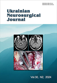
A simple CT-scan-assisted craniotomy for small superficial cortical lesions in rural conditions
DOI:
https://doi.org/10.25305/unj.298375Keywords:
craniotomy, CT scan, rural conditions, design, superficial cortical lesionAbstract
Objective: Despite the excellence and modernization in medicine and neurosurgery, many countries, including Greece, still lack neuronavigational techniques, or hospital budget to cover the neuronavigation expenses. Therefore, help in the craniotomy design is needed, not only to safely remove a superficial lesion but also to help cut the expenses of neuronavigation in cases of economic challenges. The current study aims to present a new simple technique for craniotomy design for superficial cortical lesions.
Materials and methods: The technique was applied as an urgent lifesaving method because of lacking frameless neuronavigation to 35 patients (19 males and 16 females) with superficial cortical lesions during a five-year period. This technique requires computer tomography (CT) scan, needle, and methylene blue dye. The patients were operated on at the neurosurgical department of Democritus University Hospital in Alexandroupolis, Greece.
Results: From those 35 individuals, 16 had brain metastases, six patients had meningioma, six patients had glioma tumor, two had an abscess, two patients had arteriovenous malformation (AVM) and three patients had brain hematoma. The lesion was completely resected in all the 35 patients without any complications from the craniotomy or the colorant dye infusion. The accuracy of the technique compared with the frameless neuronavigation of the literature was extremely high.
Conclusion: This is a simple and cheap technique for craniotomy design in case of superficial cortical lesions. It could be used in rural conditions or in hospitals with limited resources, as long as there is a computed tomography scan, craniotomy device and a dye stain.
References
1. Cranial approaches. General principles [Internet]. Neurosurgical atlas; 2024. https://www.neurosurgicalatlas.com/volumes/cranial-approaches/general-principles
2. Nikova A, Birbilis T. The Basic Steps of Evolution of Brain Surgery. Maedica (Bucur). 2017 Dec;12(4):297-305. [PubMed] [PubMed Central]
3. Sperati G. Craniotomy through the ages. Acta Otorhinolaryngol Ital. 2007 Jun;27(3):151-6. [PubMed] [PubMed Central]
4. Kirkpatrick DB. The first primary brain-tumor operation. J Neurosurg. 1984 Nov;61(5):809-13. [CrossRef] [PubMed]
5. Thomas NWD, Sinclair J. Image-Guided Neurosurgery: History and Current Clinical Applications. J Med Imaging Radiat Sci. 2015 Sep;46(3):331-342. [CrossRef] [PubMed]
6. Rozet I, Vavilala MS. Risks and benefits of patient positioning during neurosurgical care. Anesthesiol Clin. 2007 Sep;25(3):631-53, x. [CrossRef] [PubMed] [PubMed Central]
7. Clatterbuck R, Tamargo R: Surgical Positioning and Exposures for Cranial Procedures. In: Winn H, editor. Youmans Neurological Surgery. 5 ed. Philadelphia, Pennsylvania: Sounders Elsevier Inc, 2004: 623–645
8. Caro H. Improvement in the production of dyestuffs from methyl-aniline. US patent 204796, 1878.
9. Caro H. Verfahren zur Darstellung blauer Farbstoffe aus Dimethylanilin und anderen tertiaren aromatischen Monoaminen. German patent 1886 (December 15, 1877); British patent 3751 (October 9, 1877), 1877
10. Cooksey CJ. Quirks of dye nomenclature. 8. Methylene blue, azure and violet. Biotech Histochem. 2017;92(5):347-356. [CrossRef] [PubMed]
11. Ehrlich P: Methodologische Beiträge zur Physiologie und Pathologie der verschiedenen Formen der Leukocyten. Z Klin Med. 1880;1:553–560. https://www.pei.de/SharedDocs/Downloads/DE/institut/veroeffentlichungen-von-paul-ehrlich/1877-1885/1880-methodologisch-beitraege-physiogie-pathologie.pdf
12. Bodoni P. Dell'azione sedativa del bleu di metilene in varie forme di psicosi. Clin Med Ital. 1899;24:217–222.
13. Margetis K, Rajappa P, Tsiouris AJ, Greenfield JP, Schwartz TH. Intraoperative stereotactic injection of Indigo Carmine dye to mark ill-defined tumor margins: a prospective phase I-II study. J Neurosurg. 2015 Jan;122(1):40-8. [CrossRef] [PubMed]
14. Fogler R, Golembe E. Methylene blue injection. An intraoperative guide in small bowel resection for arteriovenous malformation. Arch Surg. 1978 Feb;113(2):194-5. [CrossRef] [PubMed]
15. Gifford SM, Peck MA, Reyes AM, Lundy JB. Methylene blue enteric mapping for intraoperative localization in obscure small bowel hemorrhage: report of a new technique and literature review: combined intraoperative methylene blue mapping and enterectomy. J Gastrointest Surg. 2012 Nov;16(11):2177-81. [CrossRef] [PubMed]
16. Liu P, Zhang H, Qiu T, Zhu W. Trans-radial artery microcatheter angiography-assisted juvenile ruptured brainstem arteriovenous malformation resection. Acta Neurochir (Wien). 2024 Jan 30;166(1):53. [CrossRef] [PubMed]
17. PROVAYBLUE (methylene blue) injection. Highlights of prescribing information [Internet]. U.S. Food and Drug Administration; 2017. https://www.accessdata.fda.gov/drugsatfda_docs/label/2017/204630s005lbl.pdf
18. Pharmacology reviews: Application number :21-670 [Internet]. Centre for drug evaluation and research; 2004. https://www.accessdata.fda.gov/drugsatfda_docs/nda/2004/21-670_VisionBlue_Pharmr.PDF
19. Evans blue [Internet]. U.S. Food and Drug Administration; 2022. https://www.accessdata.fda.gov/scripts/cder/daf/index.cfm?event=overview.process&ApplNo=008041
20. Evans JP. Warning against intrathecal use of methylene blue. Journal of the American Medical Association. 1959 Jan 31;169(5):526. doi:10.1001/jama.1959.03000220106025
21. Sharr MM, Weller RO, Brice JG. Spinal cord necrosis after intrathecal injection of methylene blue. J Neurol Neurosurg Psychiatry. 1978 Apr;41(4):384-6. [CrossRef] [PubMed] [PubMed Central]
22. ECKER A. Cerebrospinal rhinorrhea by way of the eustachian tube; report of cases with the dural defects in the middle or posterior fossa. J Neurosurg. 1947 Mar;4(2):177. [CrossRef] [PubMed]
23. Wirth D, Snuderl M, Curry W, Yaroslavsky A. Comparative evaluation of methylene blue and demeclocycline for enhancing optical contrast of gliomas in optical images. J Biomed Opt. 2014 Sep;19(9):90504. [CrossRef] [PubMed]
24. Phelan AL, Jones CM, Ceschini AS, Henry CR, Mackay DR, Samson TD. Sparing a Craniotomy: The Role of Intraoperative Methylene Blue in Management of Midline Dermoid Cysts. Plast Reconstr Surg. 2017 Jun;139(6):1445-1451. [CrossRef] [PubMed]
25. Talley Watts L, Long JA, Chemello J, Van Koughnet S, Fernandez A, Huang S, Shen Q, Duong TQ. Methylene blue is neuroprotective against mild traumatic brain injury. J Neurotrauma. 2014 Jun 1;31(11):1063-71. [CrossRef] [PubMed] [PubMed Central]
26. Shen J, Xin W, Li Q, Gao Y, Yuan L, Zhang J. Methylene Blue Reduces Neuronal Apoptosis and Improves Blood-Brain Barrier Integrity After Traumatic Brain Injury. Front Neurol. 2019 Nov 8;10:1133. [CrossRef] [PubMed] [PubMed Central]
27. Farrokhi MR, Lotfi M, Masoudi MS, Gholami M. Effects of methylene blue on postoperative low-back pain and functional outcomes after lumbar open discectomy: a triple-blind, randomized placebo-controlled trial. J Neurosurg Spine. 2016 Jan;24(1):7-15. [CrossRef] [PubMed]
28. Lee YS, Wurster RD. Methylene blue induces cytotoxicity in human brain tumor cells. Cancer Lett. 1995 Jan 27;88(2):141-5. [CrossRef] [PubMed]
29. Snuderl M, Wirth D, Sheth SA, Bourne SK, Kwon CS, Ancukiewicz M, Curry WT, Frosch MP, Yaroslavsky AN. Dye-enhanced multimodal confocal imaging as a novel approach to intraoperative diagnosis of brain tumors. Brain Pathol. 2013 Jan;23(1):73-81. [CrossRef] [PubMed] [PubMed Central]
30. Risholm P, Golby AJ, Wells W 3rd. Multimodal image registration for preoperative planning and image-guided neurosurgical procedures. Neurosurg Clin N Am. 2011 Apr;22(2):197-206, viii. [CrossRef] [PubMed] [PubMed Central]
31. Orringer DA, Golby A, Jolesz F. Neuronavigation in the surgical management of brain tumors: current and future trends. Expert Rev Med Devices. 2012 Sep;9(5):491-500. [CrossRef] [PubMed] [PubMed Central]
32. Leksell L. The stereotaxic method and radiosurgery of the brain. Acta Chir Scand. 1951 Dec 13;102(4):316-9. [PubMed]
33. Spiegel EA, Wycis HT, Marks M, Lee AJ. Stereotaxic Apparatus for Operations on the Human Brain. Science. 1947 Oct 10;106(2754):349-50. [CrossRef] [PubMed]
34. Mascott CR. In vivo accuracy of image guidance performed using optical tracking and optimized registration. J Neurosurg. 2006 Oct;105(4):561-7. [CrossRef] [PubMed]
Downloads
Published
How to Cite
Issue
Section
License
Copyright (c) 2024 Alexandrina Nikova, Efthymia Theodoropoulou, Theodossios A. Birbilis

This work is licensed under a Creative Commons Attribution 4.0 International License.
Ukrainian Neurosurgical Journal abides by the CREATIVE COMMONS copyright rights and permissions for open access journals.
Authors, who are published in this Journal, agree to the following conditions:
1. The authors reserve the right to authorship of the work and pass the first publication right of this work to the Journal under the terms of Creative Commons Attribution License, which allows others to freely distribute the published research with the obligatory reference to the authors of the original work and the first publication of the work in this Journal.
2. The authors have the right to conclude separate supplement agreements that relate to non-exclusive work distribution in the form of which it has been published by the Journal (for example, to upload the work to the online storage of the Journal or publish it as part of a monograph), provided that the reference to the first publication of the work in this Journal is included.


