Peculiarities of the cavernous cerebral malformations diagnostic and treatment
DOI:
https://doi.org/10.25305/unj.126810Keywords:
cavernous malformation, the types of clinical course and treatmentAbstract
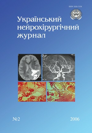
On the basis of analysis of 49 cerebral malformations observations are analyzed and the peculiarities of their diagnostic and treatment were distinguished. Observation data and results of the patients surgical treatment with microsurgical technique application substantiate the necessity of more active operative interventions usage at this complex vascular cerebral pathology treatment.
References
Вихерт Т.М., Филатов Ю.М., Шишкина Л.В. и др. Клинико-анатомическая характеристика, причины диагностических ошибок при сосудистых микромальформациях головного мозга // Нейрохирургия. — 2000. — №3. — С.3–5.
Гайдар Б.В., Парфенов В.Е., Свистов Д.В. Определение тактики хирургического лечения артериовенозных мальформаций головного мозга на основании данных минимально-инвазивного диагностического комплекса // V Междунар. симпоз. “Повреждение мозга”. — СПб, 1999. — С.311–320.
Гайтур Е.И. Сосудистые мальформации головного мозга: Автореф. дис. … канд. мед. наук. — М., 1993.
Гельфенбейн М.С., Крылов В.В. Особенности инструментальной диагностики разорвавшихся сосудистых мальформаций головного мозга // Нейрохирургия. — 2000. — №3. — С.56–60.
Евзиков Г.Ю., Крылов В.В., Новиков В.А. Кавернозная гемангиома — динамическое наблюдение или хирургическое вмешательство? // Неврол. журн. — 1998. — №4. — С.25–27.
Коновалов А.Н., Корниенко В.Н., Пронин И.Н. и др. Гематомы и скрытые сосудистые мальформации ствола мозга // Мед. визуализация. — 2001. — №2. — С.13–21.
Коновалов А.Н., Махмуров У.Б., Филатов О.М. и др. Клиника, диагностика и хирургическое лечение гематом ствола мозга // Вопр. нейрохирургии. — 1991. — №1. — С.3–6.
Медведев Ю.А., Мацко Д.Е. Аневризмы и пороки развития сосудов мозга. — СПб, 1993. — Т.2. — 140 с.
Орлов К.Ю. Кавернозные мальформации головного мозга: Автореф. дис. … канд. мед. наук. — СПб, 2003.
Панунцев В.С. Современные методы нейровизуализации в диагностике внутричерепных кавернозных мальформаций // Материалы VI Междунар. сипоз. “Современные минимально инвазивные технологии”. — СПб, 2001. — С.59–60.
Панунцев В.С., Орлов К.Ю. Диагностика внутричерепных кавернозных мальформаций, проявляющихся эписиндромом // Журн. теор. и клин. медицины. — 2000. — №3. — С.233–234.
Тютин Л.А., Яковлева Е.К. Магниторезонансная ангиография в диагностике заболеваний сосудов головы и шеи // Вестн. рентгенологии и радиологии. — 1998. — №6. — С.4–8.
Шалек Р.А., Коннов Б.А., Тютин Л.А. и др. Стереотаксическая фотонная терапия кавернозных ангиом головного мозга на отечественном линейном ускорителе электронов ЛУЭР 20-М // VII Междунар. симпоз. “Новые технологии в нейрохирургии”. — СПб, 2004. — С.141–142.
Diamond C., Torvik A., Amundsen P. Angiographic diagnosis of teleangiectasis with cavernous angioma of the posterior fossa: Report of two cases // Acta Radiol. — 1976. — V.17. — P.281–288.
Di Tulleo H.V., Stern W.E. Hemangioma calcificans: Case report of an intraparenchymatous calcified vascular hematoma with epileptogenic potential // J. Neurosurg. — 1979. — V.50. — P.110–114.
Fahlbusch R., Strauss C., Huk W. et al. Surgical removal of pontomesencephalic cavernous hemangiomas // Neurosurgery. — 1990. — V.26. — P.449–457.
Fischer E.G., Sotuel A., Welch K. Cerebral hemangioma with glial neoplasia (angioglioma): Report of two cases // J. Neurosurg. — 1982. — V.56. — P.430–434.
Kamrin R.B., Buchsbaum H.W. Large vascular malformations of the brain not visualized by serial angiography // Arch. Neurol. (Chic). — 1965. — V.13. — P.413–421.
Martin N.A., Bentson. J., Vinuella F. et al. Intraoperative digital subtraction angiography and the surgical treatment of intracranial aneurysms and vascular malformations // Ibid. — 1990. — V.73. — P.526–533.
Mizoik Н, Yoshimoto T., Suzuki J. Clinical analysis of ten cases with surgically treated brainstem cavernous angiomas // Tohoku, J. Exp. Med. — 1992. — V.166, N2. — P.259–267.
Rao V.R.K., Pillai S.M., Shenoyk T. et al. Hypervascular cavernous angioma at angiography // Neuroradiologe. — 1979. — V.79. — P.211–214.
Rapacki T.F., Brantley M.J., Farlow T.W. et al. Heterogeneity of cerebral cavernous hemangiomas diagnosed by MR imaging // J. Comput. Assist. Tomogr. — 1990. — V.14. — P.18–25.
Rigamonti D. Johnson P.C., Spetzler R.F. et al. Cavernous malformations and capillary teleangiectasia: a spectrum within a single pathological entity // Neurosurgery. — 1991. — V.28. — P.60–64.
Sakai N., Yamada H., Tanigavara T. et al. Surgical treatment of cavernous angioma involving brainstem and review of the literature // Acta Neurochir. (Vien). — 1991. — V.133. — P.138–143.
Yasargil M.G. Microneurosurgery // III B. — Stuttgard: New York: George Thieme Verlag, 1988. — P.405–435.
Zabramski J.M., Washer T.M., Spetzler R.F. et al. The natural history of familial cavernous malformations: result of an ongoing study // J. Neurosurg. — 1994. — V.80. — P.422–432.
Downloads
How to Cite
Issue
Section
License
Copyright (c) 2006 O. A. Tsimeyko, A. I. Goncharov, M. Yu. Orlov, I. I. Skorokhoda, O. G. Chernenko

This work is licensed under a Creative Commons Attribution 4.0 International License.
Ukrainian Neurosurgical Journal abides by the CREATIVE COMMONS copyright rights and permissions for open access journals.
Authors, who are published in this Journal, agree to the following conditions:
1. The authors reserve the right to authorship of the work and pass the first publication right of this work to the Journal under the terms of Creative Commons Attribution License, which allows others to freely distribute the published research with the obligatory reference to the authors of the original work and the first publication of the work in this Journal.
2. The authors have the right to conclude separate supplement agreements that relate to non-exclusive work distribution in the form of which it has been published by the Journal (for example, to upload the work to the online storage of the Journal or publish it as part of a monograph), provided that the reference to the first publication of the work in this Journal is included.


