
Mykola Pirogov’s (1810-1881) illness mystery. Computed tomography and 3D reconstruction of the head of the famous surgeon's mummy
DOI:
https://doi.org/10.25305/unj.268440Keywords:
Mykola Ivanovych Pirogov, re-embalming, computed tomography, 3D scanning, malignant tumor, cancerous ulcer of the oral cavityAbstract
Introduction. It is known that at the age of 70, the outstanding surgeon Mykola Pirogov suffered from pain and a wound of the palate on the right and had problems with eating. He was consulted by well-known doctors M.V. Sklifosovskyi, E. von Wahl, V.F. Grube, E.I. Bohdanovskyi and the famous surgeon T. Billroth, convincing him that the ulcer was benign.
On the fourth day after his death, on the initiative of his wife Baroness
O.A. von Bistrom, Pirogov's body was embalmed by permission of the church.
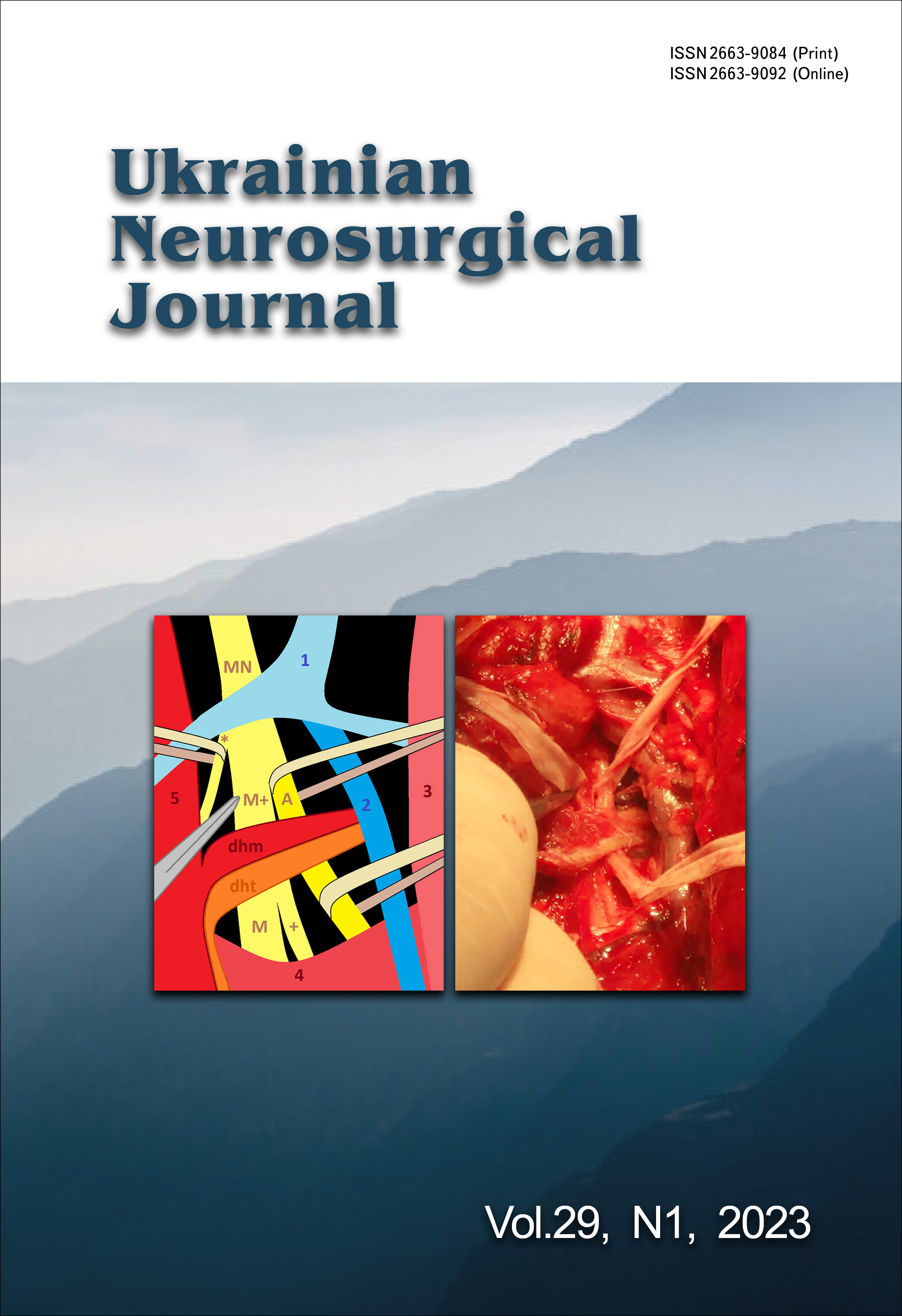
Case report. In 2018, M.I. Pirogov's body was re-embalmed in Vinnytsia according to the original method by scientists of Vinnytsia National Medical University and the National University of Life and Environmental Sciences of Ukraine.
The remains were examined using a 32-slice computer tomograph Siemens "Somatom go. Up" (Germany) with 3D reconstruction of the entire body and the head of great surgeon. According to the protocol, the slices thickness was 0.8 mm, the voltage was 110‒120 kV, the current strength was 30‒230 mA, the thickness of reconstructions was 0.8 to 3.0 mm.
Modern technologies made it possible to see destructive changes in the bones of the skull and establish the cause of Mykola Pirogov's illness and death. The 3D reconstructions prove the fact that Pirogov's diagnosis was correct. The existing bone changes indicate widespread malignancy, most likely cancer in the mouth, nasopharynx, and pterygopalatine fossa on the right.
Discussion. Natural and anthropogenic mummies are important for history and science, as they can tell us about the health conditions and lifestyle of people in the past.
Computed tomography is a non-destructive technique, and is therefore considered the gold standard for studying mummies. This method is also used during the embalming procedure and monitor the degree of preservation of the mummified body. Currently, computed tomography is widely used in mummy research to non-invasively assess the natural or anthropogenic origin, mummification embalming technique, bone and soft tissue preservation parameters, age, constitution, health status, cause of death, post-mortem injuries, etc.
Conclusions. The use of computed tomography followed by 3D reconstruction is highly likely to not only predict the future, but also shed light on the mysteries of the past.
References
1. Piombino-Mascali D, Jankauskas R, Kozakaitė J, Sutherland ML. Paleoimaging of a modern mummy from Lithuania (circa 19th-20th century). Medicina (Kaunas). 2017;53(6):410-419. [CrossRef] [PubMed]
2. Zesch S, Panzer S, Rosendahl W, Nance JW Jr, Schönberg SO, Henzler T. From first to latest imaging technology: Revisiting the first mummy investigated with X-ray in 1896 by using dual-source computed tomography. Eur J Radiol Open. 2016 Jul 25;3:172-81. [CrossRef] [PubMed] [PubMed Central]
3. Macchi V, Picardi EEE, Porzionato A, Morra A, Tabarin L, Gusella F, Grignon B, De Caro R. Friar Leopold Mandic (1866-1942): the computed tomography of the body of a saint. Surg Radiol Anat. 2018 Aug;40(8):967-975. [CrossRef] [PubMed]
4. Panzer S, Gill-Frerking H, Rosendahl W, Zink AR, Piombino-Mascali D. Multidetector CT investigation of the mummy of Rosalia Lombardo (1918-1920). Ann Anat. 2013 Oct;195(5):401-8. [CrossRef] [PubMed]
5. Villa C, Davey J, Craig PJ, Drummer OH, Lynnerup N. The advantage of CT scans and 3D visualizations in the analysis of three child mummies from the Graeco-Roman Period. Anthropol Anz. 2015;72(1):55-65. [CrossRef] [PubMed]
6. Baudouin R, Amelot A, Laprie Y, Crevier-Buchman L, Maeda S, Huynh-Charlier I, Hans S, Charlier P. Henri IV of France's larynx 3D reconstitution. Eur Arch Otorhinolaryngol. 2023 Feb;280(2):919-924. [CrossRef] [PubMed]
7. Saleem SN, Hawass Z. Computed Tomography Study of the Mummy of King Seqenenre Taa II: New Insights Into His Violent Death. Front Med (Lausanne). 2021 Feb 17;8:637527. [CrossRef] [PubMed]; [PubMed Central]
8. Saleem SN, Hawass Z. Computed Tomography Study of the Feet of Mummy of Ramesses III: New Insights on the Harem Conspiracy. J Comput Assist Tomogr. 2017 Jan;41(1):15-17. [CrossRef] [PubMed]
9. Martina MC, Cesarani F, Boano R, Fiore Marochetti E, Gandini G. Petamenophis (Padiamenemipet), an Egyptian Child Mummy Protected for Eternity: Revelation by Multidetector Computed Tomography. J Comput Assist Tomogr. 2018 Mar/Apr;42(2):178-183. [CrossRef] [PubMed]
10. Shbat A, Beránek M, Seifert Z, Kratzer J, Musil S, Klepáček I. Natural or intended mummification? Specific case of a child mummy. Anthropol Anz. 2021 Jan 5. [CrossRef] [PubMed]
11. Saleem SN, Hawass Z. Digital Unwrapping of the Mummy of King Amenhotep I (1525-1504 BC) Using CT. Front Med (Lausanne). 2021 Dec 28;8:778498. [CrossRef] [PubMed] [PubMed Central]
12. Jackowski C, Bolliger S, Thali MJ. Common and unexpected findings in mummies from ancient Egypt and South America as revealed by CT. Radiographics. 2008 Sep-Oct;28(5):1477-92. [CrossRef] [PubMed]
Downloads
Published
How to Cite
Issue
Section
License
Copyright (c) 2023 Mykola Y. Polishchuk, Oleg P. Melnyk, Ivan V. Shevchuk, Oleg P. Robak

This work is licensed under a Creative Commons Attribution 4.0 International License.
Ukrainian Neurosurgical Journal abides by the CREATIVE COMMONS copyright rights and permissions for open access journals.
Authors, who are published in this Journal, agree to the following conditions:
1. The authors reserve the right to authorship of the work and pass the first publication right of this work to the Journal under the terms of Creative Commons Attribution License, which allows others to freely distribute the published research with the obligatory reference to the authors of the original work and the first publication of the work in this Journal.
2. The authors have the right to conclude separate supplement agreements that relate to non-exclusive work distribution in the form of which it has been published by the Journal (for example, to upload the work to the online storage of the Journal or publish it as part of a monograph), provided that the reference to the first publication of the work in this Journal is included.


