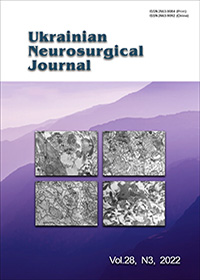
Changes in the structure of synaptic intercellular contacts in focal brain lesions
DOI:
https://doi.org/10.25305/unj.259732Keywords:
focal lesions of the brain, electron microscopy, synapseAbstract
Purpose: to evaluate changes in the structure of synaptic contacts in various types of focal brain pathology.
Materials and methods. The results of treatment of 40 cases of supratentorial focal lesions of the brain (FLB) were retrospectively evaluated. The cases are divided into groups: 30 gliomas of various degrees of malignancy and 5 consequences of TBI, 5 epilepsy. All patients underwent surgical interventions. The synaptic plasticity of axo-dendritic and axo-spiny asymmetric synapses of neurons of the VI-VII layers of the frontotemporal cortex was studied by electron microscopy. Morphometric analysis was carried out on a computer image analyzer САИ-01АВН using the software "Kappa opto-electronics GmbH" using the STATISTICA 7 program package.
The results. It was established that the density of synapses decreased in glioblastomas (GB) and craniocerebral injury (ССІ). Qualitative changes demonstrate the plasticity of architectonics of synapse, in particular due to the increase in the number of perforated synaptic contacts. Maximum thickening and diffuse stratification of the postsynaptic seal indicates a violation of the functional capacity of the postsynaptic component of the contacts. A decrease in the number of synaptic vesicles was revealed in ССІ and GB, with their rearrangement, which is probably a manifestation of synaptic dysfunction. The latter proves the irreversibility of destructive local changes and is unfavorable criterion. The risk of the formation of destructive-degenerative changes in the synaptic apparatus is 7.64 times higher in DA, 3.17 times higher in GB, and 17.31 times higher in ССІ compared to cases of epilepsy, with GB significantly increases by 13.5 times compared to DA. Therefore, the assessment of the structural features of neuroplasticity should take into account the morphogenesis of the BM in comparison with clinical data
Conclusions. In the zones of invasive growth of gliomas of various degrees of malignancy and in ССІ and epilepsy, the indicators of synaptic plasticity differ statistically significantly. The density of placement of synapses is lower in GB and ССІ. The probability of non-reversibility of destructive-degenerative changes of synapses according to the number of SVs in FLB correlates with the degree of glioma differentiation with a sensitivity of 81.0% and a specificity of 76.0%. According to the structural changes of synaptic connections in tumors, probable differences between the variants have been proven: GB and DA, the sensitivity of the discriminant model is 85.0%, the specificity is 74.0%, which is an indirect evidence of the growth rate of the tumor mass and its destructive effect on the surrounding brain matter. The obtained results are important in assessing the prognosis of the further course of the disease.
References
Turrigiano GG, Nelson SB. Homeostatic plasticity in the developing nervous system. Nat Rev Neurosci. 2004 Feb;5(2):97-107. doi: 10.1038/nrn1327
Manto M, Oulad ben Taib N, Luft AR. Modulation of excitability as an early change leading to structural adaptation in the motor cortex. J Neurosci Res. 2006 Feb 1;83(2):177-80. doi: 10.1002/jnr.20733
Horner PJ, Gage FH. Regenerating the damaged central nervous system. Nature. 2000 Oct 26;407(6807):963-70. doi: 10.1038/35039559
Chen R, Cohen LG, Hallett M. Nervous system reorganization following injury. Neuroscience. 2002;111(4):761-73. doi: 10.1016/s0306-4522(02)00025-8
Delpech JC, Madore C, Nadjar A, Joffre C, Wohleb ES, Layé S. Microglia in neuronal plasticity: Influence of stress. Neuropharmacology. 2015 Sep;96(Pt A):19-28. doi: 10.1016/j.neuropharm.2014.12.034
Wilson JT, Pettigrew LE, Teasdale GM. Structured interviews for the Glasgow Outcome Scale and the extended Glasgow Outcome Scale: guidelines for their use. J Neurotrauma. 1998 Aug;15(8):573-85. doi: 10.1089/neu.1998.15.573
Kovalenko T, Osadchenko I, Nikonenko A, Lushnikova I, Voronin K, Nikonenko I, Muller D, Skibo G. Ischemia-induced modifications in hippocampal CA1 stratum radiatum excitatory synapses. Hippocampus. 2006;16(10):814-25. doi: 10.1002/hipo.20211
Shabanov DA, Kravchenko MA. Statisticheskiy analiz dannykh v zoologii i ekologii [Internet]. Batrachos; 2011. [cited 2022 April 17]. Available from: https://batrachos.com/BioStatistica_Basis.
Rebrova OYU. Statisticheskiy analiz meditsinskikh dannykh. Primeneniye paketa prikladnykh programm STATISTICA. Moscow: MediaSfera. 2002.
Gusev YEI, Kryzhanovskiy GN. Disregulyatsionnaya patologiya nervnoy sistemy. Moscow: OOO «MIA», 2009.
Boholepova AN, Tsukanova EY. Problema neyroplastychnosty v nevrolohyy. Mizhnarodnyy nevrolohichnyy zhurnal. 2010;(8):69-72.
Johansen-Berg H, Dawes H, Guy C, Smith SM, Wade DT, Matthews PM. Correlation between motor improvements and altered fMRI activity after rehabilitative therapy. Brain. 2002 Dec;125(Pt 12):2731-42. doi: 10.1093/brain/awf282. Erratum in: Brain. 2003 Nov;126(Pt 11):2569.
Dietz V. Neuronal plasticity after a human spinal cord injury: positive and negative effects. Exp Neurol. 2012 May;235(1):110-5. doi: 10.1016/j.expneurol.2011.04.007
Hübener M, Bonhoeffer T. Neuronal plasticity: beyond the critical period. Cell. 2014 Nov 6;159(4):727-37. doi: 10.1016/j.cell.2014.10.035
Delpech JC, Madore C, Nadjar A, Joffre C, Wohleb ES, Layé S. Microglia in neuronal plasticity: Influence of stress. Neuropharmacology. 2015 Sep;96(Pt A):19-28. doi: 10.1016/j.neuropharm.2014.12.034
Froemke RC. Plasticity of cortical excitatory-inhibitory balance. Annu Rev Neurosci. 2015 Jul 8;38:195-219. doi: 10.1146/annurev-neuro-071714-034002
Griesbach GS, Hovda DA. Cellular and molecular neuronal plasticity. Handb Clin Neurol. 2015;128:681-90. doi: 10.1016/B978-0-444-63521-1.00042-X
Khan F, Amatya B, Galea MP, Gonzenbach R, Kesselring J. Neurorehabilitation: applied neuroplasticity. J Neurol. 2017 Mar;264(3):603-615. doi: 10.1007/s00415-016-8307-9
Taylor WD, MacFall JR, Payne ME, McQuoid DR, Steffens DC, Provenzale JM, Krishnan RR. Greater MRI lesion volumes in elderly depressed subjects than in control subjects. Psychiatry Res. 2005 May 30;139(1):1-7. doi: 10.1016/j.pscychresns.2004.08.004
Stogsdill JA, Eroglu C. The interplay between neurons and glia in synapse development and plasticity. Curr Opin Neurobiol. 2017 Feb;42:1-8. doi: 10.1016/j.conb.2016.09.016
Weiller C, Rijntjes M. Learning, plasticity, and recovery in the central nervous system. Exp Brain Res. 1999 Sep;128(1-2):134-8. doi: 10.1007/s002210050828
Gustin SM, Peck CC, Cheney LB, Macey PM, Murray GM, Henderson LA. Pain and plasticity: is chronic pain always associated with somatosensory cortex activity and reorganization? J Neurosci. 2012 Oct 24;32(43):14874-84. doi: 10.1523/JNEUROSCI.1733-12.2012
Dzyak LA. Kohnityvnyy ta neyrosensornyy defitsyt riznoho henezu: yak ne propustyty holovne. Ukrayinsʹkyy medychnyy chasopys.2021;(4):8-13. doi: 10.32471/umj.1680-3051.144.214545
Fleminger S, Oliver DL, Lovestone S, Rabe-Hesketh S, Giora A. Head injury as a risk factor for Alzheimer's disease: the evidence 10 years on; a partial replication. J Neurol Neurosurg Psychiatry. 2003 Jul;74(7):857-62. doi: 10.1136/jnnp.74.7.857
Mendez MF, Paholpak P, Lin A, Zhang JY, Teng E. Prevalence of Traumatic Brain Injury in Early Versus Late-Onset Alzheimer's Disease. J Alzheimers Dis. 2015;47(4):985-93. doi: 10.3233/JAD-143207
Gavett BE, Stern RA, Cantu RC, Nowinski CJ, McKee AC. Mild traumatic brain injury: a risk factor for neurodegeneration. Alzheimers Res Ther. 2010 Jun 25;2(3):18. doi: 10.1186/alzrt42
Downloads
Published
How to Cite
Issue
Section
License
Copyright (c) 2022 Viktoriia V. Vaslovytch, Artem V. Rozumenko, Leonid R. Borovyk, Anna A. Shmeleva, Volodymyr D. Rozumenko, Tetyana A. Malysheva

This work is licensed under a Creative Commons Attribution 4.0 International License.
Ukrainian Neurosurgical Journal abides by the CREATIVE COMMONS copyright rights and permissions for open access journals.
Authors, who are published in this Journal, agree to the following conditions:
1. The authors reserve the right to authorship of the work and pass the first publication right of this work to the Journal under the terms of Creative Commons Attribution License, which allows others to freely distribute the published research with the obligatory reference to the authors of the original work and the first publication of the work in this Journal.
2. The authors have the right to conclude separate supplement agreements that relate to non-exclusive work distribution in the form of which it has been published by the Journal (for example, to upload the work to the online storage of the Journal or publish it as part of a monograph), provided that the reference to the first publication of the work in this Journal is included.


