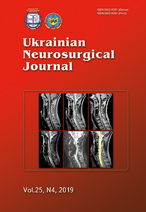
The usage of tractography of the spinal cord as a predictor of neurological disorders regression in patients with severe cervical spine and spinal cord injury
DOI:
https://doi.org/10.25305/unj.176912Keywords:
spinal cord tractography, diffusion-tensor imaging, prognosis, cervical spine injury, spinal cord lesionsAbstract
Objective: the primary assessment of the prognostic features of tractography in patients with severe subaxial cervical spine and spinal cord injury.
Materials and methods. The clinical group consisted of 5 patients admitted to the Department of Spine Surgery of Romodanov Neurosurgery Institute during the period from April to July 2019 with severe traumatic injury of the cervical spine and spinal cord. MRI was performed in the following modes: T1W, T2W, FLAIR, STIR, T2W FFE, CSF flow and DTI in 5–7 days after surgery. The dynamics of neurological disorders regression were evaluated according to the International Standards for Neurological Classification of Spinal Cord Injury. The level of spinal canal traumatic stenosis was determined by SCT both before and after surgical correction. Due to the small clinical group, statistical processing of the obtained digital indicators was not performed. The main task was to identify general patterns in order to determine the direction of further detailed studies.
Results. Spinal cord edema at the damage level was observed in 100 % of patients that corresponded to the level of neurological disorders. The presence of hemorrhagic component was detected in 3 patients, while post-traumatic spinal stenosis was: more than 50 % — in one patient, up to 50 % — one patient and less than 25 % — one patient. Spinal cord parenchyma edema was accompanied by varying degrees of compression of the cerebrospinal spaces, from subtotal to total, with the presence or absence of a hemorrhagic component that did not impact the intensity of cerebrospinal fluid flow disorders. Evaluation of DTI results showed a moderate inverse relationship between FA and MD indicators. Two patients presented with a regression of neurological disorders. Restoration of sensory-motor functions did not correlate with the degree of compression of the spinal canal or the nature of changes in the parenchyma of the spinal cord, but was noted in patients with maximum FA values and low MD.
Conclusions. Tractography is an informative predictor of the regression of neurological disorders in patients with severe spine and spinal cord injury. Further studies are needed to identify the optimal timing of its implementation and to establish diagnostically significant values of anisotropic diffusion indices.
References
1. Duane TM, Young AJ, Vanguri P, Wolfe LG, Katzen J, Han J, Mayglothling J, Whelan JF, Aboutanos MB, Ivatury RR, Malhotra AK. Defining the cervical spine clearance algorithm. J. Trauma Acute Care Surg. 2016 Sep;81(3):541-7. [CrossRef] [PubMed]
2. Kalfas I, Wilberger J, Goldberg A, Prostko RE. Magnetic Resonance Imaging in Acute Spinal Cord Trauma. Neurosurgery 1988 Sep;23(3):295-9. [CrossRef] [PubMed]
3. Mihai G, Nout YS, Tovar CA, Miller BA, Schmalbrock P, Bresnahan JC, Beattie MS. Longitudinal comparison of two severities of unilateral cervical spinal cord injury using magnetic resonance imaging in rats. J. Neurotrauma 2008 Jan;25(1):1-18. [CrossRef] [PubMed]
4. Miyanji F, Furlan JC, Aarabi B, Arnold PM, Fehlings MG. Acute Cervical Traumatic Spinal Cord Injury: MR Imaging Findings Correlated with Neurologic Outcome—Prospective Study with 100 Consecutive Patients. Radiology 2007 Jun;243(3):820-7. [CrossRef] [PubMed]
5. Rajasekaran S, Kanna RM, Shetty AP. Diffusion tensor imaging of the spinal cord and its clinical applications. J. Bone Joint Surg. Br. 2012 Aug;94-B(8):1024-31. [CrossRef] [PubMed]
6. Bosma R, Stroman PW. Diffusion tensor imaging in the human spinal cord: development, limitations, and clinical applications. Crit. Rev. Biomed. Eng. 2012;40(1):1-20. [PubMed]
7. Wang-Leandro A, Hobert MK, Kramer S, Rohn K, Stein VM, Tipold A. The role of diffusion tensor imaging as an objective tool for the assessment of motor function recovery after paraplegia in a naturally-occurring large animal model of spinal cord injury. J. Transl. Med. 2018;16(1):258. [CrossRef] [PubMed]
8. Ries M, Jones RA, Dousset V, Moonen CTW. Diffusion tensor MRI of the spinal cord. Magn. Reson. Med. 2000 Dec 1;44(6):884-92. [CrossRef]
9. Thomsen C, Henriksen O, Ring P. In vivo measurement of water self diffusion in the human brain by magnetic resonance imaging. Acta Radiol.28(3):353-61. [PubMed]
10. Alizadeh M, Intintolo A, Middleton DM, Conklin CJ, Faro SH, Mulcahey MJ, Mohamed FB. Reduced FOV diffusion tensor MR imaging and fiber tractography of pediatric cervical spinal cord injury. Spinal Cord 2017 Mar 16;55(3):314-20. [CrossRef] [PubMed]
11. Zhao C, Rao J-S, Pei X-J, Lei J-F, Wang Z-J, Yang Z-Y, Li X-G. Longitudinal study on diffusion tensor imaging and diffusion tensor tractography following spinal cord contusion injury in rats. Neuroradiology 2016 Jun 1;58(6):607-14. [CrossRef] [PubMed]
12. D’souza MM, Choudhary A, Poonia M, Kumar P, Khushu S. Diffusion tensor MR imaging in spinal cord injury. Injury 2017 Apr;48(4):880-4. [CrossRef] [PubMed]
13. Lee S, Lee YH, Chung T-S, Jeong E-K, Kim S, Yoo YH, Kim IS, Yoon C-S, Suh J-S, Park JH. Accuracy of Diffusion Tensor Imaging for Diagnosing Cervical Spondylotic Myelopathy in Patients Showing Spinal Cord Compression. Korean J. Radiol. 2015;16(6):1303. [CrossRef] [PubMed]
14. Patel SP, Smith TD, VanRooyen JL, Powell D, Cox DH, Sullivan PG, Rabchevsky AG. Serial Diffusion Tensor Imaging In Vivo Predicts Long-Term Functional Recovery and Histopathology in Rats following Different Severities of Spinal Cord Injury. J. Neurotrauma 2016;33(10):917-28. [CrossRef] [PubMed]
15. Shanmuganathan K, Gullapalli RP, Zhuo J, Mirvis SE. Diffusion Tensor MR Imaging in Cervical Spine Trauma. Am. J. Neuroradiol. 2008 Apr;29(4):655-9. [CrossRef] [PubMed]
16. Deo AA, Grill RJ, Hasan KM, Narayana PA. In vivo serial diffusion tensor imaging of experimental spinal cord injury. J. Neurosci. Res. 2006 Apr;83(5):801-10. [CrossRef] [PubMed]
17. Kelley BJ, Harel NY, Kim C-Y, Papademetris X, Coman D, Wang X, Hasan O, Kaufman A, Globinsky R, Staib LH, Cafferty WBJ, Hyder F, Strittmatter SM. Diffusion Tensor Imaging as a Predictor of Locomotor Function after Experimental Spinal Cord Injury and Recovery. J. Neurotrauma 2014 Aug 1;31(15):1362-73. [CrossRef] [PubMed]
18. Basser PJ, Jones DK. Diffusion-tensor MRI: theory, experimental design and data analysis - a technical review. NMR Biomed. 2002 Nov;15(7-8):456-67. [CrossRef] [PubMed]
19. Kim JH, Budde MD, Liang H-F, Klein RS, Russell JH, Cross AH, Song S-K. Detecting axon damage in spinal cord from a mouse model of multiple sclerosis. Neurobiol. Dis. 2006 Mar;21(3):626-32. [CrossRef] [PubMed]
20. Sun S-W, Liang H-F, Trinkaus K, Cross AH, Armstrong RC, Song S-K. Noninvasive detection of cuprizone induced axonal damage and demyelination in the mouse corpus callosum. Magn. Reson. Med. 2006 Feb;55(2):302-8. [CrossRef] [PubMed]
21. Committee Membership:, Burns S, Biering-Sørensen F, Donovan W, Graves DE, Jha A, Johansen M, Jones L, Krassioukov A, Kirshblum S, Mulcahey MJ, Read MS, Waring W. International standards for neurological classification of spinal cord injury, revised 2011. Top. Spinal Cord Inj. Rehabil. 2012 Jan;18(1):85-99. [CrossRef] [PubMed]
22. Pouw MH, van der Vliet AM, van Kampen A, Thurnher MM, van de Meent H, Hosman AJF. Diffusion-weighted MR imaging within 24 h post-injury after traumatic spinal cord injury: a qualitative meta-analysis between T2-weighted imaging and diffusion-weighted MR imaging in 18 patients. Spinal Cord 2012 Jun 7;50(6):426-31. [CrossRef] [PubMed]
23. Quencer RM, Bunge RP, Egnor M, Green BA, Puckett W, Naidich TP, Post MJ, Norenberg M. Acute traumatic central cord syndrome: MRI-pathological correlations. Neuroradiology 1992;34(2):85-94. [PubMed]
24. Demaerel P, Van Hecke P, Marchal G, Wilms G, Baert AL. MRI of intraparenchymal hematoma: responsible mechanisms. J. Belge Radiol. 1990 Aug;73(4):279-84. [PubMed]
25. Bondurant FJ, Cotler HB, Kulkarni M V, McArdle CB, Harris JH. Acute spinal cord injury. A study using physical examination and magnetic resonance imaging. Spine (Phila. Pa. 1976). 1990 Mar;15(3):161-8. [PubMed]
26. Cotler HB, Kulkarni M V, Bondurant FJ. Magnetic resonance imaging of acute spinal cord trauma: preliminary report. J. Orthop. Trauma 1988;2(1):1-4. [PubMed]
27. SCHAEFER DM, FLANDERS A, NORTHRUP BE, DOAN HT, OSTERHOLM JL. Magnetic Resonance Imaging of Acute Cervical Spine Trauma. Spine (Phila. Pa. 1976). 1989 Oct;14(10):1090-5. [CrossRef] [PubMed]
28. Schaefer DM, Flanders AE, Osterholm JL, Northrup BE. Prognostic significance of magnetic resonance imaging in the acute phase of cervical spine injury. J. Neurosurg. 1992 Feb;76(2):218-23. [CrossRef] [PubMed]
29. Talbott JF, Whetstone WD, Readdy WJ, Ferguson AR, Bresnahan JC, Saigal R, Hawryluk GWJ, Beattie MS, Mabray MC, Pan JZ, Manley GT, Dhall SS. The Brain and Spinal Injury Center score: a novel, simple, and reproducible method for assessing the severity of acute cervical spinal cord injury with axial T2-weighted MRI findings. J. Neurosurg. Spine 2015 Oct;23(4):495-504. [CrossRef] [PubMed]
30. Sun L-Q, Shen Y, Li Y-M. Quantitative magnetic resonance imaging analysis correlates with surgical outcome of cervical spinal cord injury without radiologic evidence of trauma. Spinal Cord 2014 Jul 6;52(7):541-6. [CrossRef] [PubMed]
31. Wilson JR, Grossman RG, Frankowski RF, Kiss A, Davis AM, Kulkarni A V, Harrop JS, Aarabi B, Vaccaro A, Tator CH, Dvorak M, Shaffrey CI, Harkema S, Guest JD, Fehlings MG. A clinical prediction model for long-term functional outcome after traumatic spinal cord injury based on acute clinical and imaging factors. J. Neurotrauma 2012 Sep;29(13):2263-71. [CrossRef] [PubMed]
32. Heinemann AW, Michael Linacre J, Wright BD, Hamilton BB, Granger C. Measurement characteristics of the Functional Independence Measure. Top. Stroke Rehabil. 1994 Sep 16;1(3):1-15. [CrossRef] [PubMed]
33. Osterthun R, Tjalma TA, Spijkerman DCM, Faber WXM, van Asbeck FWA, Adriaansen JJE, Post MWM. Functional independence of persons with long-standing motor complete spinal cord injury in the Netherlands. J. Spinal Cord Med. 2018 Aug 20;1-8. [CrossRef] [PubMed]
34. Skinner NP, Lee S-Y, Kurpad SN, Schmit BD, Muftuler LT, Budde MD. Filter-probe diffusion imaging improves spinal cord injury outcome prediction. Ann. Neurol. 2018 Jul;84(1):37-50. [CrossRef] [PubMed]
35. Fehlings MG, Martin AR, Tetreault LA, Aarabi B, Anderson P, Arnold PM, Brodke D, Burns AS, Chiba K, Dettori JR, Furlan JC, Hawryluk G, Holly LT, Howley S, Jeji T, Kalsi-Ryan S, Kotter M, Kurpad S, Kwon BK, Marino RJ, Massicotte E, Merli G, Middleton JW, Nakashima H, Nagoshi N, Palmieri K, Singh A, Skelly AC, Tsai EC, Vaccaro A, Wilson JR, Yee A, Harrop JS. A Clinical Practice Guideline for the Management of Patients With Acute Spinal Cord Injury: Recommendations on the Role of Baseline Magnetic Resonance Imaging in Clinical Decision Making and Outcome Prediction. Glob. Spine J. 2017 Sep 5;7(3_suppl):221S-230S. [CrossRef] [PubMed]
36. Gattozzi D, Yekzaman B, Jack M, O’Bryan M, Arnold P. Early ventral surgical treatment without traction of acute traumatic subaxial cervical spine injuries. Surg. Neurol. Int. 2018;9(1):254. [CrossRef] [PubMed]
37. Ramu J, Herrera J, Grill R, Bockhorst T, Narayana P. Brain fiber tract plasticity in experimental spinal cord injury: diffusion tensor imaging. Exp. Neurol. 2008 Jul;212(1):100-7. [CrossRef] [PubMed]
38. Wrigley PJ, Gustin SM, Macey PM, Nash PG, Gandevia SC, Macefield VG, Siddall PJ, Henderson LA. Anatomical Changes in Human Motor Cortex and Motor Pathways following Complete Thoracic Spinal Cord Injury. Cereb. Cortex 2009 Jan 1;19(1):224-32. [CrossRef] [PubMed]
39. Sun P, Murphy R, Gamble P, George A, Song S-K, Ray W. Diffusion Assessment of Cortical Changes, Induced by Traumatic Spinal Cord Injury. Brain Sci. 2017 Feb 17;7(12):21. [CrossRef] [PubMed]
40. Ellingson BM, Kurpad SN, Schmit BD. Functional correlates of diffusion tensor imaging in spinal cord injury. Biomed. Sci. Instrum. 2008;44:28-33. [PubMed]
41. Kim JH, Loy DN, Wang Q, Budde MD, Schmidt RE, Trinkaus K, Song S-K. Diffusion Tensor Imaging at 3 Hours after Traumatic Spinal Cord Injury Predicts Long-Term Locomotor Recovery. J. Neurotrauma 2010 Mar;27(3):587-98. [CrossRef] [PubMed]
42. Koskinen E, Brander A, Hakulinen U, Luoto T, Helminen M, Ylinen A, Öhman J. Assessing the State of Chronic Spinal Cord Injury Using Diffusion Tensor Imaging. J. Neurotrauma 2013 Sep 15;30(18):1587-95. [CrossRef] [PubMed]
43. Ellingson BM, Salamon N, Hardy AJ, Holly LT. Prediction of Neurological Impairment in Cervical Spondylotic Myelopathy using a Combination of Diffusion MRI and Proton MR Spectroscopy. PLoS One 2015 Oct 2;10(10):e0139451. [CrossRef] [PubMed]
44. Tetreault L, Kopjar B, Nouri A, Arnold P, Barbagallo G, Bartels R, Qiang Z, Singh A, Zileli M, Vaccaro A, Fehlings MG. The modified Japanese Orthopaedic Association scale: establishing criteria for mild, moderate and severe impairment in patients with degenerative cervical myelopathy. Eur. Spine J. 2017 Jan 24;26(1):78-84. [CrossRef] [PubMed]
45. Gu M, Gao Z, Li X, Guo L, Lu T, Li Y, He X. Conditioned medium of olfactory ensheathing cells promotes the functional recovery and axonal regeneration after contusive spinal cord injury. Brain Res. 2017 Jan 1;1654(Pt A):43-54. [CrossRef] [PubMed]
46. BASSO DM, BEATTIE MS, BRESNAHAN JC. A Sensitive and Reliable Locomotor Rating Scale for Open Field Testing in Rats. J. Neurotrauma 1995 Feb;12(1):1-21. [CrossRef] [PubMed]
47. Jirjis MB, Valdez C, Vedantam A, Schmit BD, Kurpad SN. Diffusion tensor imaging as a biomarker for assessing neuronal stem cell treatments affecting areas distal to the site of spinal cord injury. J. Neurosurg. Spine 2017 Feb;26(2):243-51. [CrossRef] [PubMed]
48. Li XF, Yang Y, Lin CB, Xie FR, Liang WG. Assessment of the diagnostic value of diffusion tensor imaging in patients with spinal cord compression: a meta-analysis. Brazilian J. Med. Biol. Res. = Rev. Bras. Pesqui. medicas e Biol. 2016 Jan;49(1):e4769. [CrossRef] [PubMed]
Downloads
Published
How to Cite
Issue
Section
License
Copyright (c) 2019 Ievgenii I. Slynko, Oleksii S. Nekhlopochyn, Kristiana O. Robak

This work is licensed under a Creative Commons Attribution 4.0 International License.
Ukrainian Neurosurgical Journal abides by the CREATIVE COMMONS copyright rights and permissions for open access journals.
Authors, who are published in this Journal, agree to the following conditions:
1. The authors reserve the right to authorship of the work and pass the first publication right of this work to the Journal under the terms of Creative Commons Attribution License, which allows others to freely distribute the published research with the obligatory reference to the authors of the original work and the first publication of the work in this Journal.
2. The authors have the right to conclude separate supplement agreements that relate to non-exclusive work distribution in the form of which it has been published by the Journal (for example, to upload the work to the online storage of the Journal or publish it as part of a monograph), provided that the reference to the first publication of the work in this Journal is included.


