
Analysis of stereotactic biopsy results and magnetic resonance imaging and histological observations of brain lesions
DOI:
https://doi.org/10.25305/unj.130893Keywords:
focal disease of the brain, stereotactic biopsy, magnetic resonance imagingAbstract
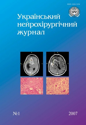
Stereotactic biopsy, using MRI data was performed in 38 patients with tumors and other brain lesions. MRI was made in standard success according special protocol with cut off thickness3 mm. Most difficult diagnostic and technical moments were tumor knots, necrotic areas, their geterogenesis and few samples for research. The results of stereotactic biopsy were analyzed and their neurosurgical, morphological and radiological aspects were considered.
References
Combs S.E., Widmer V., Thilmann C., Hof H. Stereotactic radiosurgery (SRS): treatment option for recurrent glioblastoma multiforme (GBM) // Cancer. — 2005. — V.104, N10. — P.2168–2173.
Ferreira M.P., Pereira Filho A.D. et al. Stereotactic computed tomography-guided brain biopsy: diagnostic yield based on a series of 170 patients // Surg. Neurol. — 2006. — V.65, suppl.1. — P.27–32.
Heper A.O., Erden E., Savas A., Ceyhan K. An analysis of stereotactic biopsy of brain tumors and nonneoplastic lesions: a prospective clinicopathologic study // Surg. Neurol. — 2005. — V.64, suppl.2. — P.82–88.
Woodworth G., McGirt M.J., Samdani A. et al. Accuracy of frameless and frame-based image-guided stereotactic brain biopsy in the diagnosis of glioma: comparison of biopsy and open resection specimen // Neurol. Res. — 2005. — V.27, N4. — P.358–362.
Downloads
Published
How to Cite
Issue
Section
License
Copyright (c) 2007 O. Yu. Chuvashova, A. B. Gryazov, K. R. Kostyuk, T. A. Malysheva

This work is licensed under a Creative Commons Attribution 4.0 International License.
Ukrainian Neurosurgical Journal abides by the CREATIVE COMMONS copyright rights and permissions for open access journals.
Authors, who are published in this Journal, agree to the following conditions:
1. The authors reserve the right to authorship of the work and pass the first publication right of this work to the Journal under the terms of Creative Commons Attribution License, which allows others to freely distribute the published research with the obligatory reference to the authors of the original work and the first publication of the work in this Journal.
2. The authors have the right to conclude separate supplement agreements that relate to non-exclusive work distribution in the form of which it has been published by the Journal (for example, to upload the work to the online storage of the Journal or publish it as part of a monograph), provided that the reference to the first publication of the work in this Journal is included.


